样品制备有哪些注意事项?(Western Blot Analysis, Western Blotting, WB)
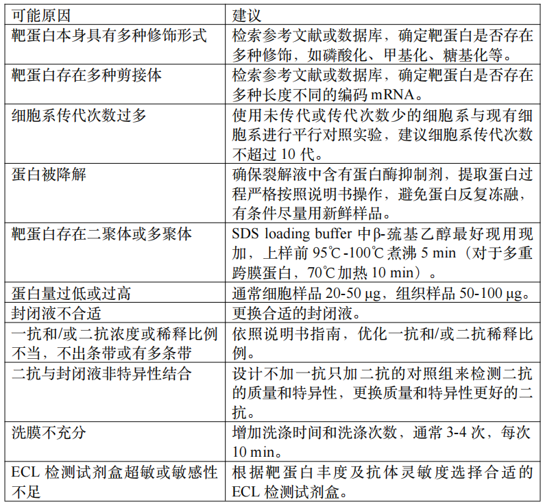
使用干净、无菌的器具,避免外源性污染;
选择合适的裂解液和裂解方法,以确保充分裂解而不破坏蛋白结构;
通过离心去除细胞碎片、核酸、脂质等杂质,只保留上清液中的蛋白质,注意离心速度和时间,避免蛋白损失或聚集;
获得的蛋白样品分装保存于-20°C或-80°C,避免反复冻融;
样品上样SDS-PAGE分析前,煮样品时防止爆管。
使用干净、无菌的器具,避免外源性污染;
选择合适的裂解液和裂解方法,以确保充分裂解而不破坏蛋白结构;
通过离心去除细胞碎片、核酸、脂质等杂质,只保留上清液中的蛋白质,注意离心速度和时间,避免蛋白损失或聚集;
获得的蛋白样品分装保存于-20°C或-80°C,避免反复冻融;
样品上样SDS-PAGE分析前,煮样品时防止爆管。


In Western blotting, if a band at the expected size can be detected by an antibody in wild-type (WT) cell lysates but disappears or significantly diminishes in gene-silenced cell lysates, the antibody is considered to be specific. Similarly, if an antibody can stain the WT cells (as indicated by strong fluorescent signals), but the signals are either lost or significantly reduced in gene-silenced cells, this antibody is deemed specific in immunofluorescence (IF) or flow cytometry (F) applications. Based on our criteria, an average of 30% antibodies tested are specific for use in Western blotting. When it comes to immunocytochemistry and immunofluorescence (IC-IF) or flow cytometry (F), only 20% of antibodies are specific.
Most SARS-CoV-2 monoclonal antibodies are developed using recombinant N or S proteins. These antibodies will react with the recombinant proteins; however, they may not react with true viruses. For instance, we developed 8 monoclonal anti-S antibodies using recombinant S proteins, only 1 of which worked with SARS-CoV-2 and human specimens. We have validated our antibodies using real coronavirus and human nasopharyngeal swabs in different applications so that you can be sure that these antibodies work without trying and error.
In Western blotting, if a band at the expected size can be detected by an antibody in wild-type (WT) cell lysates but disappears or significantly diminishes in gene-silenced cell lysates, the antibody is considered to be specific. Similarly, if an antibody can stain the WT cells (as indicated by strong fluorescent signals), but the signals are either lost or significantly reduced in gene-silenced cells, this antibody is deemed specific in immunofluorescence (IF) or flow cytometry (F) applications. Based on our criteria, an average of 30% antibodies tested are specific for use in Western blotting. When it comes to immunocytochemistry and immunofluorescence (IC-IF) or flow cytometry (F), only 20% of antibodies are specific.
Since the beginning of the COVID-19 pandemic, a large number of monoclonal and polyclonal antibodies against SARS-CoV-2 N and S proteins have entered the antibody market, most of which have been validated with recombinant proteins. There is no doubt that these antibodies react with their respective antigens, however, whether they work with the actual virus or human samples still need to be tested. In our experience, only 15% of antibodies that reacted with their respective antigens functioned well with SARS-CoV-2 and human specimens.
At certain critical points, we strongly recommend using our reagents. Our experiences show that certain proteins in the whole cell lysate are at picogram and even femtogram levels. Detecting these proteins in Western blotting using regular ECL reagents is a seemingly impossible task. In this case, our custom PiQ (Cat#636) or FeQ (Cat#226) ECL reagents can detect picogram- and femtogram-level proteins, respectively. Second, we recommend you always use our FadeStop fluorescence mounting medium (Cat#270) or FadeStop fluorescence mounting medium with DAPI (Cat#272, to counterstain nuclei) in immunofluorescent staining. These antifade fluorescence mounting media have passed our testing and can preserve fluorescence after two months when stored at -20°C or after as many as twelve laser scans using a confocal microscope. Finally, we recommend using our IntactProteinTM Cell Lysis Kit (Cat#415) to extract whole cell lysates from cultured mammalian cells. This kit is particularly formulated to maintain the integrity as well as the signal moiety (such as phosphorylation) of all sizes of proteins. Since this kit avoids sonication, it protects proteins, particularly large-sized proteins (> 100 kDa), from fragmentation.
Yes, but we cannot guarantee that it will work. Our antibodies are only tested for use in Western blotting (WB), immunocytochemistry and immunofluorescence (IC-IF), and flow cytometry (F) applications. However, it is highly probable that these validated antibodies can be used in other applications.